Common Retinal Diseases
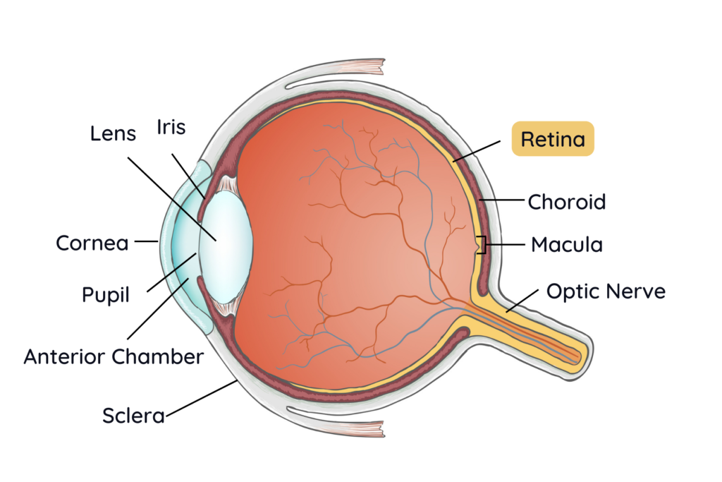
The retina is the delicate layer found in the back of your eye that functions like the film in a camera, capturing and sending visual signals to your brain from the outside world. There are numerous diseases that can affect the retina and result in vision loss. Diabetic retinopathy and macular degeneration are among the most common retinal diseases requiring treatment, and separate pages have been dedicated to these diseases. Two other common retinal conditions, retinal detachment and retinal vein and artery occlusions are briefly described below in our FAQ.
More Retinal Disease Resources
Common Retinal Diseases FAQ
A retinal detachment occurs when the retina is pulled away from the back part of the eye. This can be either a partial or complete detachment. When this occurs, the retina is no longer able to be supplied with the nutrients it needs and becomes permanently damaged without prompt treatment.
The most common form of retinal detachment occurs when a tear develops in the retina and fluid passes through the opening and pushes the retina up off the back of the eye. A retinal tear may develop for several reasons. The most common reason is a result of aging, when the vitreous gel separates from the retina and pulls on it hard enough to create a tear. Other risk factors include being very near-sighted, having a family history of retinal detachment, previous retinal detachments or tears and previous trauma or eye surgery.
Those who are high-risk should be aware of the symptoms of retinal tears and detachment, which include:
- Sudden appearance of many floaters
- Seeing flashing lights
- Change or loss in peripheral vision that moves toward central vision
Retinal detachment is often a medical emergency. Without immediate treatment, it can cause irreversible damage and vision loss. See an eye professional as soon as possible if you are concerned this may be happening.
There are several surgical methods used to treat a retinal tear or detachment, including laser photocoagulation, vitrectomy, scleral buckle and pneumatic retinopexy. Our expert ophthalmologists who specialize in these treatments come from the Vitreo-Retinal Consultants group in Wichita, Kansas. Our partnership with their practice allows patients to receive prompt diagnosis and treatment.
A retinal vein occlusion (RVO) occurs when the retinal veins that carry blood away from your retina become blocked, typically by a blood clot or the buildup of cholesterol. Two types of RVO are central retinal vein occlusions (CRVO) and branch retinal vein occlusions (BRVO). The type depends on which vein is affected. Patients with either types of RVO may suffer from gradual changes in vision, a sudden loss of vision, or no symptoms at all. Retinal swelling, bleeding and even eye pain from high eye pressure can result, ultimately leading to vision loss.
A retinal artery occlusion occurs when a retinal artery that carries blood to your retina becomes blocked. The symptoms of a blocked retinal artery usually occur suddenly and lead to temporary or permanent vision loss in a single part or all of your visual field. Retinal artery occlusions are a type of stroke and considered a medical emergency. Delayed medical treatment of issues affecting the retinal blood vessels increases the risk of retinal artery occlusion.
Both types of occlusions are caused by a blockage in either your retinal veins or arteries, so good cardiovascular health is important in preventing these conditions. People with high blood pressure, cardiovascular disease, high cholesterol, tobacco use, diabetes and obesity are at greater risk of developing a blockage.
With both retinal vein and artery occlusions there is a great risk for irreversible vision loss and accurate diagnosis and prompt treatment is important. If you are experiencing changes in vision, such as sudden vision loss or blind spots call your physician to be seen as soon as possible.
CRVO:
BRVO:
There are few effective treatments available for helping vision loss in retinal artery occlusions. More treatment options are available for retinal vein occlusions, which are mostly aimed at stopping leaking blood vessels. Examples of these treatments for RVO include anti-VEGF medication, a class of drugs that are injected into your eye and laser photocoagulation. Repeat treatments may be necessary to prevent further leakage of blood vessels and damage.
Our partnership with the expert ophthalmologists of Vitreo-Retinal Consultants Group from Wichita, Kansas allows patients to get treatment promptly and in many non-emergent cases, this care can be provided in our Garden City clinic by these visiting physicians.
Schedule Your Visit
Our doctors are here to help address your concerns about your eyes and are experts in finding solutions that work best for you.